
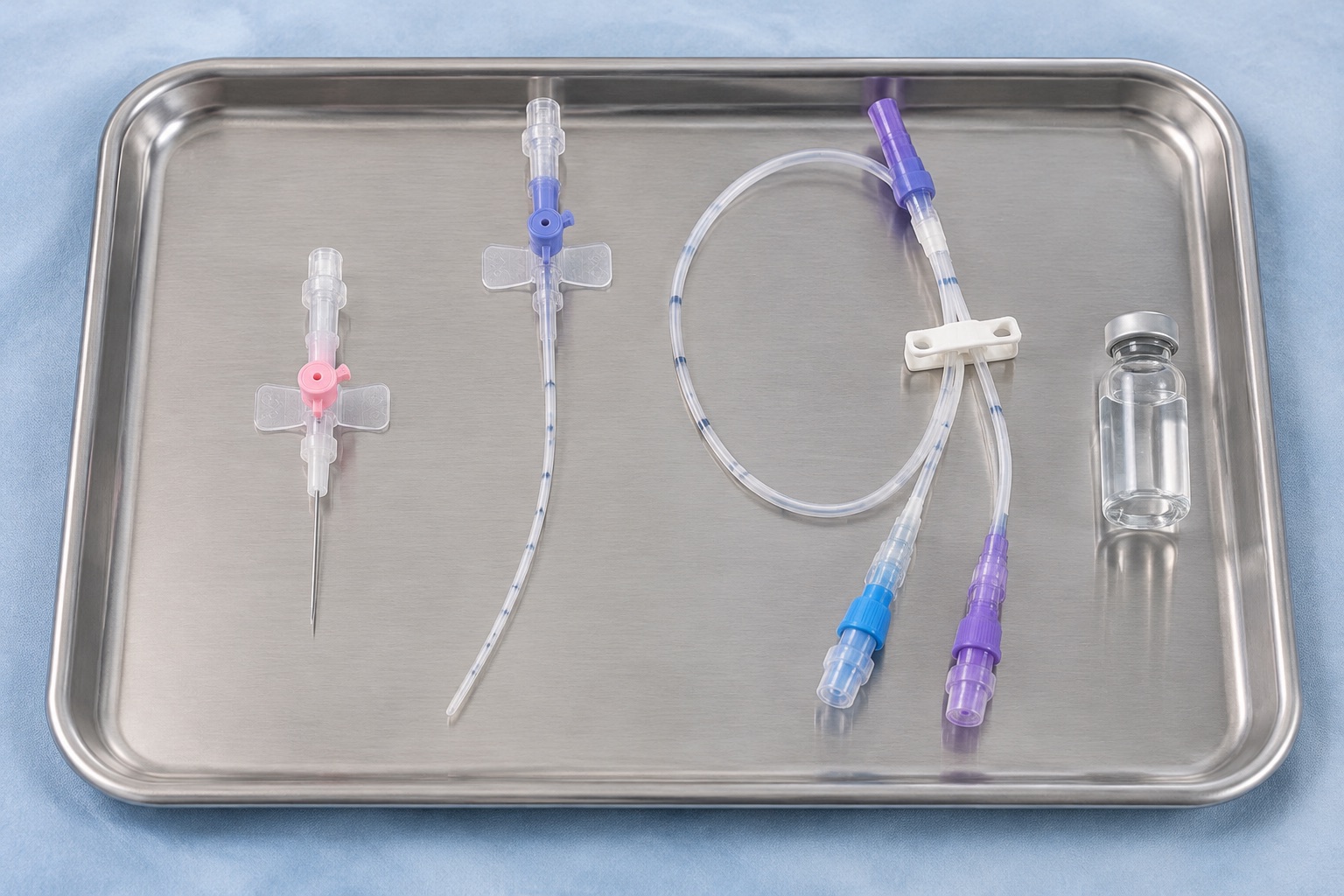
1. Science-Backed Vascular Access Selection: Aligning INS 2024 and MAGIC Criteria
Selecting the wrong vascular access device (VAD) at admission triggers a predictable cascade of preventable patient harm: repeated painful venipunctures, chemical thrombophlebitis, delayed antimicrobial dosing, catheter-related bloodstream infection (CLABSI), and upper-extremity deep vein thrombosis (DVT). For decades, inpatient wards defaulted to placing peripherally inserted central catheters (PICCs) whenever peripheral intravenous access felt inconvenient, exposing short-stay patients to unnecessary central-line complications. Modern science-backed infusion protocols have ended that ad-hoc practice by uniting two complementary evidence engines: the Infusion Nurses Society (INS) 2024 Infusion Therapy Standards of Practice (9th Edition) (Nickel et al., Journal of Infusion Nursing 2024) and the Michigan Appropriateness Guide for Intravenous Catheters (MAGIC) (Chopra et al., Annals of Internal Medicine 2015).
Rather than treating catheter choice as an isolated nursing task, both frameworks require a structured five-variable bedside assessment before any needle touches skin: (1) chemical profile of every ordered infusate (vesicant status, osmolarity, pH, and irritant index), (2) anticipated therapy duration across inpatient and post-discharge settings, (3) concurrent infusion schedule and Y-site compatibility requirements, (4) patient-specific vascular anatomy and renal preservation constraints (including chronic kidney disease stage 3b to 5 vein preservation for future arteriovenous fistulas), and (5) care setting capabilities for sterile dressing and flush maintenance.
Institutions that embed this decision logic into computerized provider order entry (CPOE) consistently reduce inappropriate PICC insertions by 25% to 40% while eliminating hazardous peripheral vesicant runs. For the standard-by-standard regulatory breakdown behind these rules, consult our INS 2024 Standards of Practice Crosswalk and our CDC CLABSI Prevention Bundle.
2. Physicochemical Decision Boundaries: Osmolarity (>900 mOsm/L), pH Extremes, and Vesicant Chemistry
Blood plasma maintains a tightly buffered physiological equilibrium at 285 to 295 mOsm/L and pH 7.35 to 7.45. When a parenteral solution deviates sharply from those coordinates inside a small-caliber forearm vein (where local blood flow averages only 20 to 40 mL/min), fluid shifts osmotically out of vascular endothelial cells, causing cellular shrinkage, inflammatory cytokine release, fibrin sheath formation, and acute chemical thrombophlebitis. By contrast, placing the catheter tip in the lower third of the superior vena cava (SVC) at the cavoatrial junction delivers the infusate into roughly 2,000 mL/min of turbulent blood flow, diluting hyperosmolar or acidic chemistry within milliseconds.
Earlier infusion standards enforced a rigid 600 mOsm/L peripheral ceiling and strictly prohibited any solution with pH below 5.0 or above 9.0. Beginning with the 2021 revision (Gorski, Home Healthcare Now 2021) and refined in the INS 2024 9th Edition, evidence shifted toward a more nuanced three-tier biochemical envelope:
- Tier 1 Peripheral-Compatible Zone (<600 mOsm/L, pH 5.0 to 9.0, Non-Vesicant): Isotonic crystalloids (0.9% NaCl at 308 mOsm/L, Lactated Ringer's at 273 mOsm/L), most cephalosporins (such as cefazolin and ceftriaxone), and dilute standard electrolytes infuse safely through short peripheral catheters (PIVC) or midline catheters without accelerated endothelial injury.
- Tier 2 Caution and Dilution Zone (600 to 900 mOsm/L or Mild Irritants): Peripheral parenteral nutrition formulated under 900 mOsm/L, 10% dextrose (505 mOsm/L, acidic pH ~4.5), or moderately concentrated potassium chloride (up to 40 mEq/L in large peripheral veins) carries escalating phlebitis risk. Protocols permit short-term peripheral delivery only through a large-caliber proximal forearm vein with hourly site inspection, maximum dilution, and immediate escalation to central access if tenderness, erythema, or induration develops.
- Tier 3 Mandatory Central Venous Access (>900 mOsm/L, Vesicants, or Severe pH Extremes): Solutions above 900 mOsm/L (central TPN exceeding 12.5% dextrose, 3% hypertonic sodium chloride at 1,026 mOsm/L, concentrated calcium or sodium bicarbonate infusions) and non-cytotoxic or cytotoxic vesicants (continuous vasopressors beyond short emergent rescue, intravenous amiodarone, phenytoin at pH 12.0, anthracyclines, vinca alkaloids) demand central venous access terminating at the cavoatrial junction. Notably, INS 2024 stresses that low pH alone does not fully predict necrosis: vancomycin (pH ~3.9, osmolarity ~291 mOsm/L) is an intense endothelial irritant that can be diluted to <=5 mg/mL for very brief (<=5 day) peripheral therapy only when strictly monitored, whereas longer vancomycin courses or vesicant agents must transition to central access to prevent severe extravasation tissue injury.
3. Peripheral IV vs. Midline vs. PICC: Comparative Clinical Matrix and Real-World Complication Rates
One of the highest-impact clinical discoveries in modern vascular access research comes from the 48-hospital Michigan Hospital Medicine Safety Consortium cohort analyzing 10,863 catheters (5,105 midlines vs. 5,758 PICCs) placed for short-term indications such as difficult venous access or intravenous antibiotics (Swaminathan et al., JAMA Internal Medicine 2022; commentary in Auron, Annals of Internal Medicine 2022). Patients receiving a midline catheter experienced less than half the rate of major complications compared with matched patients receiving a PICC (1.5% vs. 3.2%; adjusted odds ratio 1.99 favoring midlines), driven by a four-fold reduction in bloodstream infections (0.4% vs. 1.6%) and a more than three-fold drop in catheter occlusion (2.1% vs. 7.0%), with comparable or lower symptomatic DVT rates (1.0% vs. 1.6%).
That evidence underpins the core INS 2024 and MAGIC operational matrix below. Note the absolute clinical red line separating midlines from PICCs: because a midline tip terminates in the peripheral axillary/basilic vein rather than the superior vena cava, a midline is never a substitute for a central line when infusing vesicants or >900 mOsm/L solutions, even for 48 hours.
| Vascular Access Device | Target Dwell & Tip Location | Osmolarity, pH & Vesicant Rules | Y-Site & Blood Draw Capability | Benchmark Complication & Preservation Profile |
|---|---|---|---|---|
| Short Peripheral IV (PIVC) 20G to 24G (1 to 1.75 in) |
<= 5 days (clinically indicated replacement per INS 2024). Tip in superficial forearm/hand vein. | < 600 mOsm/L preferred (absolute max 900 mOsm/L with intensive monitoring); pH 5 to 9; no continuous vesicants. | Single lumen only. Avoid Y-site co-infusion of incompatible drugs; flush with 0.9% NaCl between boluses. Routine blood draws discouraged. | Lowest BSI risk (~0.1%), but 35% to 50% all-cause failure rate (infiltration, dislodgement, phlebitis). First-line for short inpatient stays. |
| Extended-Dwell Peripheral (ED-PIVC) 18G to 22G (2.5 to 3.5 in) |
Up to 7 to 14 days under ultrasound guidance. Tip in deep forearm or brachial/cephalic vein below axilla. | Strictly peripherally compatible solutions only (<= 900 mOsm/L, non-vesicant, non-continuous irritant). | Single lumen; high flow rate capability for CT power injection if rated; occasional blood draw only if institutional policy allows. | Bridges DIVA patients for 5 to 10 day stays without central line risk; requires sterile ultrasound sleeve and catheter-to-vein ratio <= 45%. |
| Midline Catheter 3 Fr to 5 Fr (3 to 8 in) |
6 to 14 days (up to 28 days for select compatible OPAT). Tip at or below axillary line in basilic, cephalic, or brachial vein. | Peripheral device rules apply: strictly prohibit vesicants, chemotherapy, TPN >900 mOsm/L, and continuous vancomycin (>5 days). | Single or dual lumen; incompatible Y-site drugs still converge in low-flow peripheral vein, so separate timing remains essential. | JAMA Intern Med 2022: 1.5% major complications, 0.4% BSI, 2.1% occlusion, 1.0% DVT. Preferred over PICC for compatible <= 14-day courses. |
| PICC Line (Central) 4 Fr single / 5 Fr dual |
> 14 days to 6 months (or any duration for central-only infusates). Tip confirmed at cavoatrial junction (SVC) via ECG or X-ray. | Permits >900 mOsm/L TPN, extreme pH, vesicants, and continuous chemotherapy due to rapid 2,000 mL/min central hemodilution. | Multi-lumen options allow simultaneous incompatible infusions via independent exit ports; reliable serial laboratory blood sampling. | JAMA Intern Med 2022: 3.2% major complications, 1.6% CLABSI, 7.0% occlusion, 1.6% DVT. Avoid in CKD stage 3b to 5 without nephrology approval. |
| Implanted Venous Port Titanium / Polysulfone |
Months to years (intermittent cyclic access). Subcutaneous reservoir accessed with non-coring Huber needle; tip at cavoatrial junction. | Gold standard for intermittent vesicant oncology chemotherapy, cyclic biologics, and long-term hyperosmolar infusions. | Single or dual-chamber models; closed subcutaneous barrier permits showering/swimming when de-accessed. | Lowest CLABSI rate among long-term central devices (~0.1 to 0.2 per 1,000 catheter-days) and highest outpatient quality of life. |
For home care maintenance once a central line is selected, review our bedside maintenance protocols for PICC Line SASH Flushing and Sterile Dressings and Implanted Port Non-Coring Huber Needle Access.
4. Y-Site Compatibility, Lumen Architecture, and Catheter-to-Vein Ratio Physics (≤45% Rule)
A frequent driver of unnecessary multi-lumen central lines is unplanned Y-site drug incompatibility. When two parenteral medications meet inside a shared administration set or single-lumen catheter dead space, rapid pH shifts can precipitate insoluble microcrystals within seconds (for example, ceftriaxone co-infused with calcium-containing solutions, phenytoin meeting dextrose, or piperacillin-tazobactam colliding with aminoglycosides or vancomycin). Yet ordering a triple-lumen PICC reflexively to solve scheduling conflicts violates both MAGIC and INS 2024 Standard 26.
Why does every extra lumen carry a steep hemodynamic penalty? Expanding from a 4-French single-lumen PICC (1.33 mm outer diameter) to a 6-French triple-lumen PICC (2.00 mm outer diameter) more than doubles the cross-sectional area obstructing the basilic or brachial vein. Under INS Standard 26, the catheter outer diameter must not exceed 45% of the native vein internal diameter measured without a tourniquet on ultrasound. That means a 4 Fr catheter requires a minimum vein diameter of 3.0 mm, a 5 Fr dual-lumen catheter requires at least 3.7 mm, and a 6 Fr triple-lumen catheter requires at least 4.5 mm. Violating the 45% catheter-to-vein threshold slows perivascular laminar flow, activating Virchow's triad and multiplying symptomatic upper-extremity DVT risk up to four-fold.
Pharmacy & Nursing 4-Step Lumen Minimization Protocol:
- 1. Audit Real-Time Y-Site Compatibility First: Cross-check every active IV medication against an updated clinical compatibility reference (Trissel's or Micromedex) before requesting a second lumen. Review high-fatality precipitation pairs in our Intravenous Drug Incompatibility Guide.
- 2. Stagger Intermittent Schedules: Convert overlapping q8h or q12h antimicrobial infusions to staggered administration windows separated by a 10 mL preservative-free 0.9% NaCl pulsatile flush, allowing a single-lumen catheter to handle multiple agents safely.
- 3. Separate Continuous High-Alert Agents by Clinical Class: Dedicate one lumen exclusively to parenteral nutrition (TPN) or continuous vasopressors; never piggyback incompatible boluses into a TPN or vasoactive carrier line.
- 4. Respect Nephrology Vein Preservation Boundaries: In patients with eGFR <45 mL/min/1.73 m2 (CKD stage 3b to 5), MAGIC strongly marks PICCs and upper-extremity midlines as inappropriate due to subclavian stenosis and loss of future hemodialysis fistula vessels; consult nephrology for a small-bore tunneled internal jugular catheter instead.
5. Bedside Escalation Protocol: Difficult Vascular Access (DIVA), Ultrasound Guidance, and Daily Necessity Audits
Even the most rigorous written algorithm fails if frontline nurses attempt four or five blind peripheral sticks before escalating. INS 2024 Standard 28 codifies a hard clinical escalation limit: no clinician should perform more than two unsuccessful peripheral cannulation attempts, and total attempts per patient should not exceed four before activating a vascular access specialist with real-time bedside ultrasound.
Patients presenting with validated Difficult Intravenous Access (DIVA) indicators, such as history of multiple prior hospitalizations, obesity, chronic edema, chemotherapy-sclerosed veins, or absence of visible and palpable veins, should bypass blind attempts altogether and receive first-pass ultrasound-guided cannulation using an extended-dwell peripheral catheter (ensuring at least 50% to 65% of the catheter shaft resides inside the vein lumen to prevent premature dislodgement). Pairing ultrasound vessel-depth mapping with the 45% catheter-to-vein ratio check lifts first-pass insertion success above 85% while cutting hematoma formation and arterial puncture risk.
Finally, science-backed vascular access stewardship does not end on insertion day. Every morning during multidisciplinary rounds, clinicians must execute a three-question daily catheter necessity audit aligned with CDC HICPAC and INS 2024 standards: (1) Can intravenous antimicrobials, electrolytes, or analgesics be stepped down to high-bioavailability oral therapy today? (2) If IV access remains required, does the current regimen still meet central-line criteria, or can a PICC be de-escalated before discharge? (3) Are insertion site dressings clean, dry, intact, and free of erythema, tenderness, or fibrin-sheath sluggishness? Removing an idle central catheter 48 hours earlier eliminates cumulative daily CLABSI risk far more reliably than any antimicrobial coating alone.